The Relationship Between Dye Structure and Its Use in Expansion Microscopy
Expansion Microscopy (ExM) relies on the physical enlargement of biological samples to achieve nanoscale resolution using conventional light microscopes. A critical component of this process is the use of fluorescent dyes, which must remain anchored and functional within the expanded matrix to provide reliable and high-resolution imaging. The chemical structure of these dyes plays a pivotal role in their performance and compatibility with ExM protocols.
Key Considerations for Dye Structure in ExM
Molecular Stability
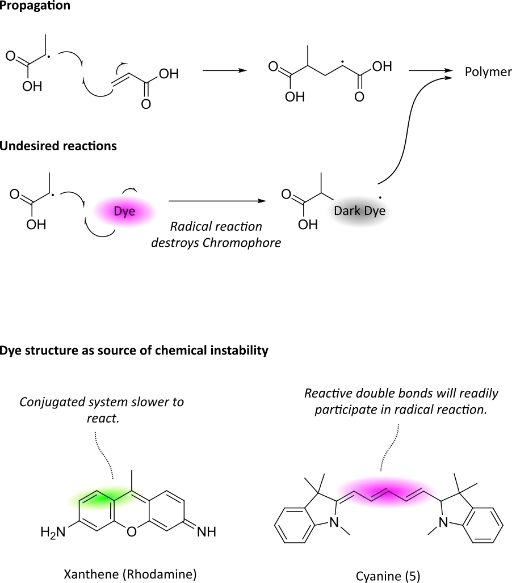
Dyes used in ExM must withstand the harsh chemical treatments required during sample preparation, such as enzymatic digestion and hydrogel embedding. Structures that are resistant to photobleaching and chemical degradation ensure signal retention throughout the process.
Spectral Properties: The dye’s excitation and emission spectra should be chosen based on the imaging setup and potential overlap with other fluorescent labels. Optimizing spectral properties minimizes crosstalk and ensures clear, multicolor imaging of expanded samples. The spectral characteristics of fluorescent dyes can shift upon binding to biomolecules, with changes in emission intensity, peak wavelength, or fluorescence lifetime often observed. Such effects arise from alterations in the dye’s local environment, including polarity, charge distribution, and proximity to quenching groups within proteins, nucleic acids, or membranes. In some cases, binding can reduce brightness or introduce spectral shifts that complicate imaging. However, in Expansion Microscopy, these issues are frequently mitigated by the decrowding effect: as the sample expands, biomolecules are physically separated within the hydrogel, reducing intermolecular quenching and self-absorption. This spatial reorganization often results in increased dye brightness and more stable spectral properties, ultimately improving the clarity and reliability of fluorescence readouts.
Dye Preference: The affinity of fluorescent dyes for biological targets is strongly influenced by their chemical structure. Hydrophobic dyes tend to insert into lipid bilayers, making them effective for membrane labeling, while aromatic or intercalating dyes often show high affinity for nucleic acids. Similarly, reactive functional groups can drive preferential association with proteins through covalent or noncovalent interactions. Charged dyes, in particular, display striking localization patterns: cationic dyes are known to accumulate in mitochondria, driven by the organelle’s negative membrane potential, while anionic or pH-sensitive dyes may preferentially associate with lysosomes or other acidic compartments. These intrinsic affinities can be exploited for targeted staining, but in Expansion Microscopy workflows, they also require careful consideration to ensure specificity and balanced distribution across sample structures.
In Expansion Microscopy, high fluorescence intensity is crucial, as signals become diluted across the physically enlarged sample volume. The brightness of a dye is determined by the product of its molar absorption coefficient (how efficiently it absorbs light) and its quantum yield (the fraction of absorbed photons re-emitted as fluorescence). Dyes with quantum yields approaching 100% are most common in the blue to green-yellow range, where shorter conjugated systems efficiently emit light. As dyes are redshifted into the orange to far-red regions, quantum yields typically decrease, a consequence of the need for longer conjugated systems to achieve absorption at these wavelengths. However, these extended dye structures also exhibit higher molar absorption coefficients, which can partially compensate for the lower quantum yield. This balance between absorption and emission efficiency makes careful dye selection critical for ensuring bright, reliable signals in expanded samples.