Cytoskeleton Imaging
Examine the networks that shape cells. Explore labelling and anchoring chemistry for a closer view of cytoskeletal organisation in expanded samples.
A closer look at
cellular architecture
Cytoskeletal networks help shape cells, organise their contents and support movement and division. Imaging their arrangement brings individual filaments, bundles and wider networks into view.
Expansion microscopy physically separates retained labels within a swellable hydrogel, making closely spaced features easier to distinguish with fluorescence microscopy. For cytoskeletal imaging, the challenge is to carry the spatial information from the labelled network through sample processing into the expanded gel.
Target-specific probes with anchoring functionality connect these two steps: recognising the structure of interest and retaining its labelling for imaging.

Why choose Chrometra?
Anchoring built in
Cytoskeleton probes combine target recognition with a polymerisable group for incorporation into the expansion gel.
Direct target recognition
Phalloidin-based and taxane-derived probes recognise F-actin and polymerised tubulin without a primary antibody.
Flexible detection formats
Choose from fluorescent probes and formats for later detection to suit your labelling sequence and imaging channels.
Reagents for cytoskeleton imaging
Explore target-binding chemistry, detection formats and fluorescent variants.
Explore related applications
See cellular organisation from another perspective, with approaches for studying membrane architecture and protein localisation.
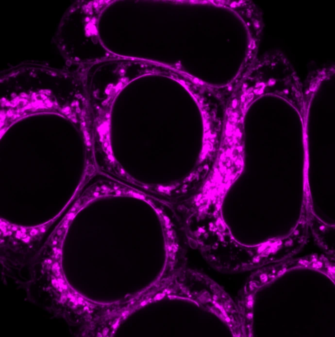
Membrane Imaging
Explore membrane organisation through lipid-based labelling, providing a different view of cellular architecture.
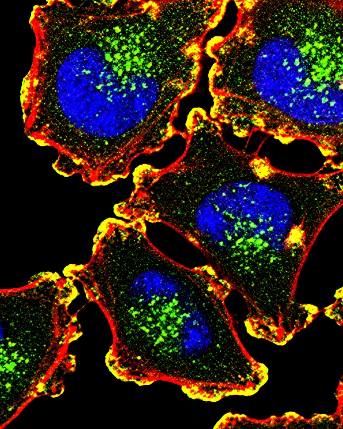
Protein & Antibody Labelling
Investigating a specific protein? Explore chemistry for labelling and anchoring antibodies directed against your target.
Let's talk about your experiment
Tell us about your work and what you’d like to achieve. We’re here to answer your labelling questions and help you choose the right reagents. We'd love to hear from you!