Membrane Imaging
Trace the boundaries that organise cells. Explore lipid-based labelling and anchoring chemistry for studying membrane organisation in expanded samples.
A closer look at cellular boundaries
Membranes define the cell’s outer boundary and organise its internal compartments. Imaging their arrangement provides a view of cellular architecture, revealing the outlines and relationships between membrane-rich structures.
In expansion microscopy, carrying that information into the expanded sample requires a label that can remain attached to the hydrogel during processing. Functionalised lipid probes address this by inserting into biological membranes and introducing a polymerisable group for gel incorporation. The retained labelling provides a record of membrane organisation after expansion.
The resulting image depends on the lipid probe, its fluorescent label and the preparation of the sample. Selecting these together helps establish the membrane contrast needed for your imaging question.

Why choose Chrometra?
Anchoring built in
A polymerisable group incorporates the probe into the hydrogel, helping retain membrane-associated labelling through subsequent processing
A choice of lipid formats
Phospholipid and sphingolipid options, with different fluorescent variants, allow selection around your sample and imaging channels.
Membrane-directed labelling
Functionalised lipid probes insert into biological membranes, providing structural contrast through lipid-based labelling.
Reagents for membrane imaging
Find lipid-based probes by membrane-labelling functionality and fluorescent variant.
Explore related applications
Place membrane organisation in a wider cellular context, from supporting cytoskeletal networks to the distribution of glycan-rich structures.
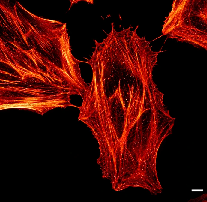
Cytoskeleton Imaging
Examine the filament networks that help shape cells and organise their contents, with labelling approaches for actin and microtubules.
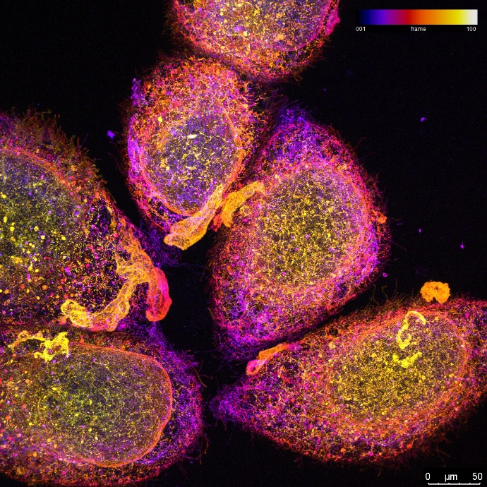
Glycan Imaging
Investigate carbohydrate-rich structures with lectin-based labelling, offering another perspective on cell surfaces and intracellular organisation.
Let's talk about your experiment
Tell us about your work and what you’d like to achieve. We’re here to answer your labelling questions and help you choose the right reagents. We'd love to hear from you!